CT Scan

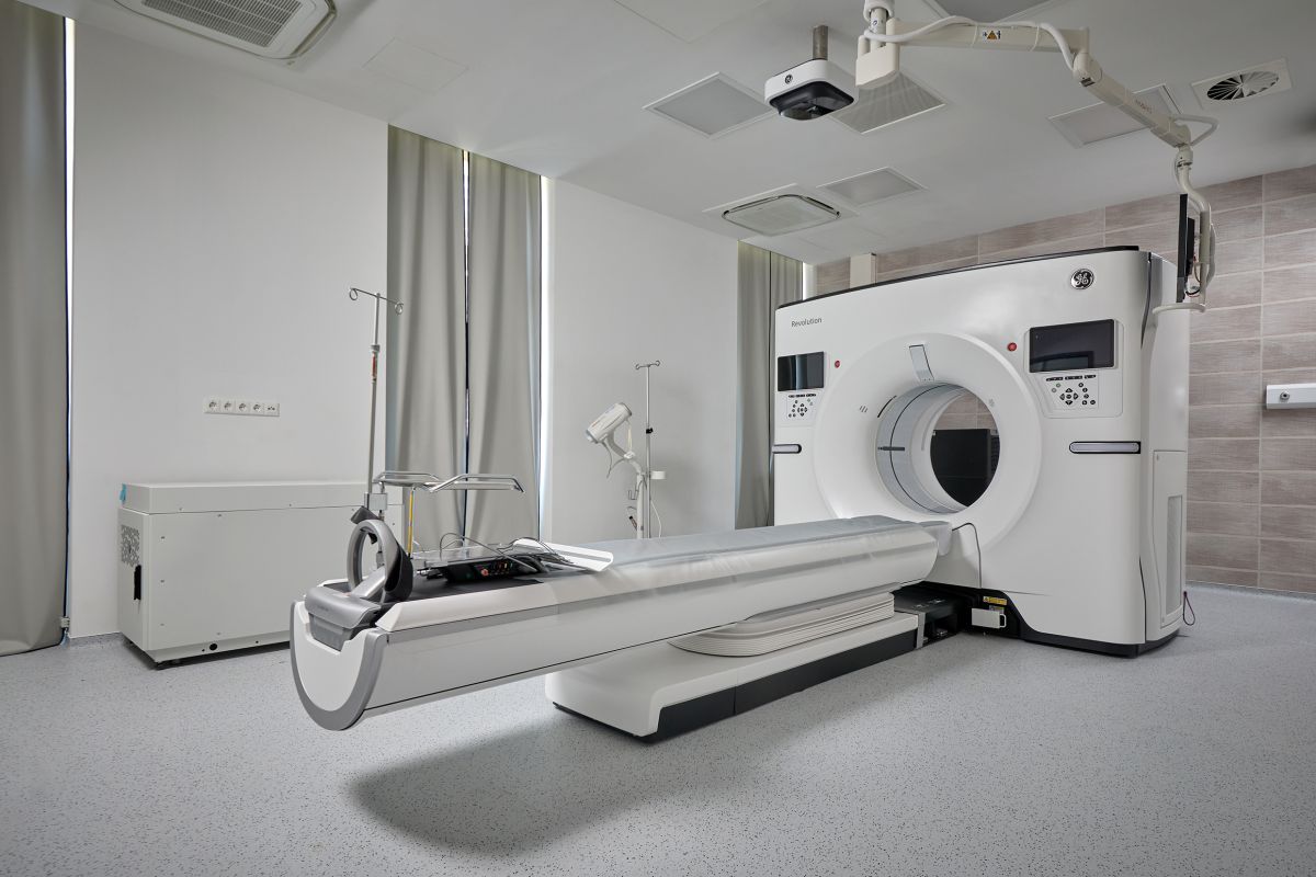
At our clinic – uniquely in Hungary – we perform necessary examinations using the state-of-the-art, 256-slice General Electric CT Revolution Apex, a cutting-edge CT system designed to provide the best possible image quality for every patient with the lowest achievable radiation dose. It is an efficient tool in medical imaging, combining innovation and functionality to meet the demands of modern healthcare professionals. The system is extremely fast, capturing full-body images in seconds, significantly reducing scan time.

What is a CT scan?
A CT (Computed Tomography) scan uses X-rays to create a series of detailed images of specific areas of the body, allowing the visualization of bones and internal organs. CT can provide answers to questions that other diagnostic methods (MRI, ultrasound, X-ray) may not fully address.

During a CT scan, the X-ray tube rotates around the body part being examined, while a detector on the opposite side captures the X-rays passing through the tissues. A computer then processes these signals to create detailed images. Different tissues absorb X-rays differently, and if tissue composition changes due to a pathological process, its X-ray absorption also changes. This makes abnormalities, tumors, organ injuries, bleeding, and inflammation visible on the images.

What are the advantages of the examination?

  • Fast and painless
  • Provides precise, accurate images of anatomical structures
  • No feeling of confinement, suitable for claustrophobic patients
  • Can be performed on patients with pacemakers, implanted metal, or prostheses
  • Low-risk imaging method

When is contrast material required?

Depending on the clinical question, a non-contrast (native) scan may be sufficient, but some examinations can only be performed with contrast. Contrast helps visualize vessels and soft tissues more clearly and allows precise determination of the type, location, and extent of abnormalities.

Contrast is an iodine-containing compound and is administered via a vein. Before the scan, a cannula is inserted into a vein in the arm or hand, through which the contrast is delivered during the examination.

Rarely, contrast may cause a hypersensitivity reaction, usually mild. For this reason, it is important to arrive fasting for a contrast-enhanced CT, avoiding food for at least 4 hours before the scan to reduce potential nausea.

Contrast administration requires adequate kidney function. A recent eGFR lab result (within 1 month) is necessary.

Patients taking metformin should suspend the medication 48 hours before the scan and may resume it after the scan depending on lab results.

Since the contrast is excreted by the kidneys, drink plenty of fluids after the scan to help flush it from the body.

When is a CT scan recommended?

A CT scan is recommended when other imaging methods cannot provide a clear diagnosis.

As CT involves radiation, it is performed only with an official medical referral or after a specialist consultation, clearly defining the body region and the need for contrast.

Screening CT scans without referral

Low-dose native chest CT indications:

  • Family history of lung cancer
  • High-risk current or former smokers
  • Chronic lung diseases (COPD)
  • Unclear findings on chest X-ray

Cardiac CT – Calcium score:
Evaluates coronary artery calcifications to assess cardiovascular risk. Our 256-slice CT can capture the heart between two beats, providing high-resolution images without beta-blockers, and eliminating motion blur.

Indications:

  • Early coronary artery calcification risk assessment
  • Family history of heart attack
  • Unexplained chest pain
  • Moderate cardiovascular risk


When is the scan not performed?

  • Pregnancy: CT is not performed
  • Young patients: Ultrasound or MRI is preferred to avoid radiation exposure

Duration of the CT scan

  • Native CT: 5–10 minutes (depending on type)
  • Contrast CT: 15–25 minutes

Preparation for the scan

  • Native CT requires no special preparation
  • For contrast CT: fasting 4–6 hours before the scan
  • Abdominal or pelvic scans must be performed on an empty stomach, as the gastrointestinal tract is examined after ingesting contrast fluid

When are results available?

The images are evaluated by a radiologist specialized in CT, usually within 3 working days.

  • Reports are sent by email and available on the EESZT platform
  • Image data is provided on CD/DVD immediately after the scan

Body regions examined by CT

Musculoskeletal diseases and sports injuries:

  • Spine and limb CT scans
  • Image-guided minimally invasive interventions (e.g., small joint inflammation)
  • CT arthrography after ultrasound-guided contrast injection for:
  • Shoulder: rotator cuff injuries
  • Hip: cartilage damage
  • Knee: meniscus and cartilage defects
  • Wrist: ligament injuries

Cardiology:

  • Heart CT: motion-free imaging at high resolution for any heart rate
  • Coronary CT: evaluates coronary arteries, myocardial perfusion, and cardiac function after contrast
  • Calcium score: native heart CT for coronary calcification risk assessment

Oncology:

  • Chest–abdomen–pelvis CT for cancer staging
  • Low-dose techniques allow imaging of entire organs or systems, including follow-up exams

Neurology:

  • Stroke evaluation with full-brain perfusion and CT angiography
  • Early diagnosis of cerebral ischemia, aneurysms, stenosis, and tumors

Internal medicine:

  • Imaging of the gastrointestinal system with native and contrast CT

Angiology:

  • Carotid and peripheral arterial CT angiography for aneurysms, stenosis, calcifications, plaques, congenital anomalies, vascular injuries, and thrombus detection


Dr. Rudas Gábor

MR Specialist Manager

Rudas G Oldalra

Contact

Looking forward to meet you.

1024 Budapest, Lövőház utca 39. I. em. bal oldali ajtó

E-mail info@mind.hu
Opening hours
Mon-Fri 8:00 - 18:00
Sat 8:00 - 12:00 (all even weeks native MR)
Sun closed